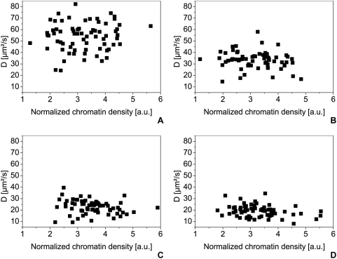
Figure 10. Diffusion coefficients of the eGFP-oligomers in HEK293 cells.
The diffusion coefficients in HEK293 cells are plotted against the normalized chromatin density given by the H2A-mRFP1 fluorescence intensity for the eGFP-monomer (A) (70 samples), the dimer (B) (63 samples), the trimer (C) (66 samples) and the tetramer (D) (67 samples). All measurements were carried out at 37°C in 5% CO2 atmosphere, using a water immersion objective with a NA of 1.2.

