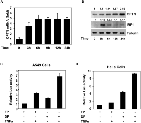
Figure 1. Induction of optineurin gene expression and promoter activation by TNFα.
(A), A549 cells were treated with TNFα (10 ng/ml) for indicated time (3–24 hours). Total RNA was then isolated and the level of optineurin mRNA was determined by real time RT-PCR. GAPDH was used as a control. (B) Effect of TNFα on optineurin protein level. A549 cells were treated with TNFα (10 ng/ml) for indicated time. The cell lysates were then prepared for immunoblotting which was performed using antibodies against optineurin, IRF-1 and tubulin (loading control). The numbers at the top indicate relative amount of protein. Activation of optineurin promoter by TNFα in A549 cells (C) or HeLa cells (D). Cells grown in 24 well plates were transfected with 100 ng of optineurin promoter-reporter plasmid (full length construct pGL-FP or deletion construct pGL-DP) along with pCMV.SPORT β-gal plasmid. After 6 hours of transfection TNFα was added (10 ng/ml). After another 18 hours cell lysates were prepared for reporter assays. Luciferase activities relative to untreated control (taken as 1.0) are shown (n = 3) after normalizing with β-galactosidase enzyme activities.

