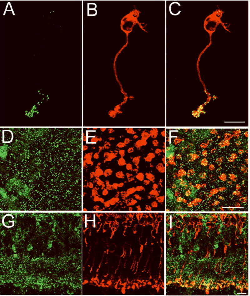
Figure 1. An isolated rod bipolar cell contains ≈ 34 synaptic ribbons.

Confocal fluorescence images of a freshly isolated mouse rod bipolar cell (A-C), a whole mount retinal section (D-F) in the lower sublamina of the inner plexiform layer (IPL) and a retinal vertical section (G-I) were double-labeled for CtBP2/ribeye (green, A, D,G) and PKC-α (red, B, E, H). CtBP2/ribeye marks the synaptic ribbon, while PKC-α labels rod bipolar cells. CtBP2/ribeye immunofluorescence (green) is prominent in the synaptic terminals of isolated rod bipolar cells and in the IPL, the retinal layer in which the terminals of all bipolar cells reside. The number of synaptic ribbons in a rod bipolar cell was determined from the number of CtBP2/ribeye puncta that resided within the terminal cluster of an individual isolated PKC-α positive bipolar cell (34 ± 4; n=10). Scale bars represent 10 μm.
