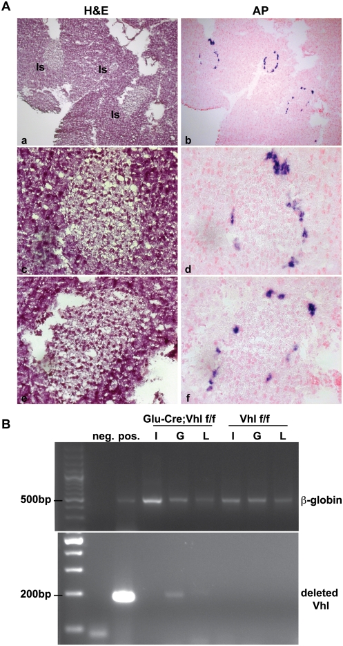
Figure 1. Analysis of Glu-Cre;Vhl colony.
A. Validation of the Glu-Cre transgenic line with Z/AP reporter mice. H&E (panels a, c, e) and alkaline phosphatase (AP; panels b, d, f) staining of a representative Glu-Cre;Z/AP pancreas at 5 months of age. Expression of Glu-Cre localized in α-cells at the outer ring of endocrine islets, as indicated by purple AP stain. Pancreatic islets (Is) are as indicated in panel a, and images are shown at 100× (a, b) and 400× (c, d, e, f) magnification. B. Genotyping PCR using genomic DNA isolated from different cell populations of the endocrine pancreas in Glu-Cre;Vhl f/f and Vhl f/f mice at 27 months of age. Insulin-positive β-cells (I), glucagon-positive α-cells (G), and lectin-positive endothelial cells (L) were collected via flow cytometry. Negative (neg.) and positive (pos.) controls are shown next to the ladder (far left lane). Top panel shows the PCR for β-globin alleles to demonstrate the presence of genomic DNA for each cell population. Bottom panel indicates the presence of deleted Vhl alleles only in α-cells of Glu-Cre;Vhl f/f pancreas.

