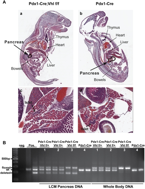
Figure 3. Histological and molecular analyses of control and Pdx1-Cre;Vhl f/f pup pancreas.
A. H&E staining of representative Pdx1-Cre, Vhl f/f (panels a and c) and control Pdx1-Cre (panels b and d) mouse pups at postnatal day 3 (P3). Magnified (50×) pup pancreas are shown (panels c and d), and islets are indicated by arrows. B. PCR analysis of Vhl allele status. DNA isolated from whole pup section (whole body DNA) and from pancreas via laser capture microdissection (LCM pancreas DNA) was used to detect Vhl allele status (floxed, wildtype-wt, deleted). Genotyping PCR was performed in duplicate for each pup (#1–#4). PCR results for Pdx1-Cre;Vhl f/f (#3)and Pdx1-Cre (#4) are the same mouse pups shown in A.

