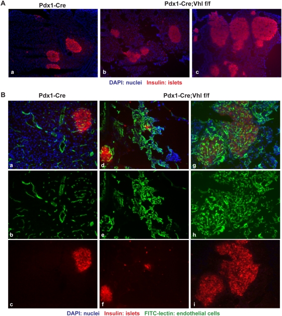
Figure 5. Histological analysis of the endocrine pancreas in Pdx1-Cre;Vhl f/f mice.
A. Immuno-fluorescent staining of representative pancreas in Pdx1-Cre (panel a) and Pdx1-Cre;Vhl f/f mice (panels b and c) to demonstrate the abnormally shaped and hyperplastic islets (red) in Pdx1-Cre;Vhl f/f mice at 16–18 months of age. Images are taken at 100×. B. Immuno-fluorescent images of representative pancreas in Pdx1-Cre (panels a–c) and Pdx1-Cre;Vhl f/f (panels d–i) to demonstrate hypervascularity within islets of Pdx1-Cre;Vhl f/f mice. Blood vessels are visualized via FITC-lectin injection (green; panels b, e, and h) while pancreatic islets are identified using an anti-insulin antibody (red; panels c, f, and i). Images are taken at 200×.

