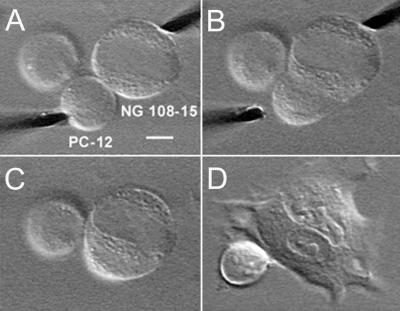
Figure 3.
PC 12 x NG 108–15 heterokaryon formation. The NG 108–15 cells are growing on the glass surface (processes out of focus), and PC 12 cells were added from suspension. In A, the cells are shown during ac dielectrophoresis (1–1.8 kV/cm, 1 MHz, for 1 min). Dielectrophoresis and electrofusion were performed in a hypoosmotic sorbitol fusion medium. (B) Twenty seconds after the dc fusion pulse (1.8 kV/cm, 1 ms), the PC 12 cell appears to be fused to the NG 108–15 cell, but the resulting membrane of the heterokaryon is not fully reorganized. After about 40 s, the fusion of the two cells appears to be complete (C). After growth in cell-culture media for 20 h in incubator (37°C, 90% humidity and 5% CO2 atmosphere), the heterokaryon developed new processes and grows on the glass surface (D). The third cell (also a PC 12 cell) (Upper Left), which is attached to the PC 12 cell, remains unaffected throughout the fusion process. In the first three panels, it is slightly swollen from the hypoosmotic fusion media. Bar = 10 μm.

