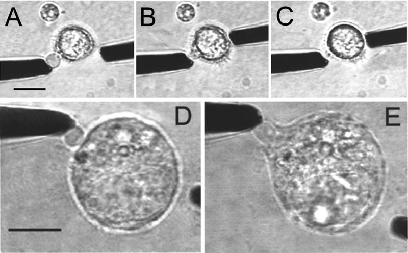
Figure 4.
Bright-field images taken (A) before, (B) during, and (C) after the electrofusion (≈8 kV/cm, 4 ms) of a PC vesicle (Left) with a Cos 7 cell (Right) in Hepes-buffered saline solution. To assist vesicle–cell electrofusion, 1.25% dimethyl sulfoxide and 20% Milli-Q-water was added to the external buffer solution. D–E show fusion or hemifusion between a NG 108–15 cell (protease-treated for 30 min) and a PC vesicle with incorporated γ-GT. The resulting fluorescence (not shown) from the vesicle, after fusion, was too weak to detect by the CCD camera used for these experiments. The fusion protocol was the same as in A–C. In E, the microelectrode is gently pulled away from the cell, and the vesicle (which is attached to the microelectrode) is stretched but does not detach from the cell. Bar = 10 μm.

