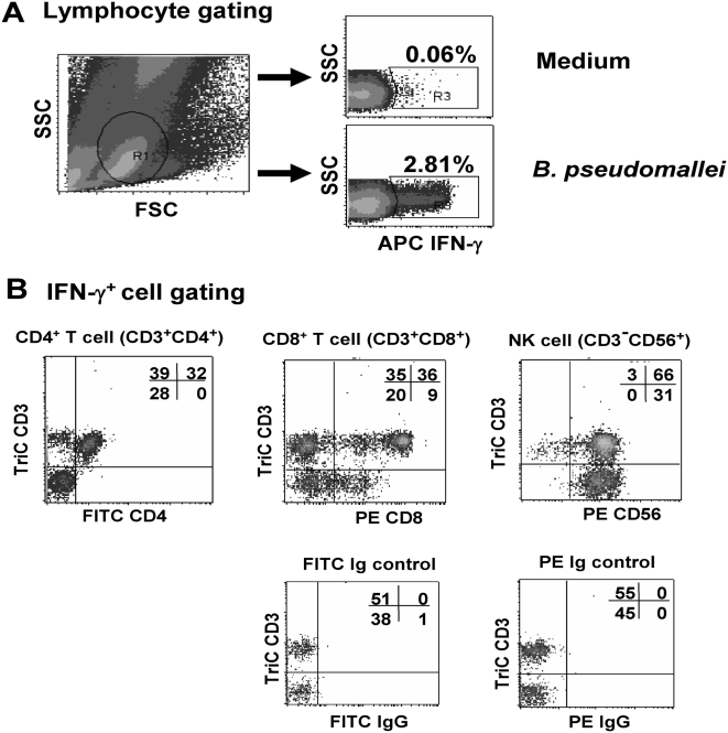
Figure 4. Identification of IFN-γ secreting T cells responding to whole B. pseudomallei by four-color flow cytometry.
Whole blood samples from eight recovered melioidosis cases and six seropositive control subjects were incubated with whole B. pseudomallei for 12 hours and stained for intracellular IFN-γ vs. three immune cell surface markers (Tri-color anti-CD3, FITC-anti-CD4 and PE-anti-CD8 or PE-anti-CD56). The profile from one representative donor of seropositive control subjects, (A) gated on lymphocyte cells, and (B) gated on IFN-γ+ cells.

