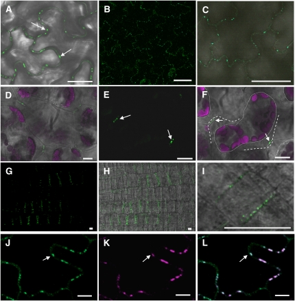
Figure 2.
YFP-PDCB1 Shows Targeting to Pds.
(A) Transgenic expression of 35SproYFP-PDCB1 in Arabidopsis showing fluorescence as punctate spots on the walls of epidermal pavement cells.
(B) and (C) Transgenic expression of PDCB1pro:YFP-PDCB1 in Arabidopsis showing similar patterns of fluorescence as in (A), in the absence of overexpression.
(D) to (F) Similar patterns of fluorescence for YFP-PDCB1 were seen in spongy mesophyll cells where the fluorescent punctae were restricted to regions of wall–wall contact between adjacent cells and were stably located following plasmolysis; (F) is the same as (E) except with combined differential interference contrast microscopy. Arrows indicate YFP-PDCB1 fluorescence on adjoining walls. Dotted lines indicate periphery of retracted protoplast and dashed lines the position of the cell wall. Fluorescence is shown for GFP in green and chlorophyll autofluorescence in magenta.
(G) to (I) YFP-PDCB1 labeling on new anticlinal division walls in newly divided root epidermal cells immediately behind the root meristem.
(J) to (L) Colocalization (e.g. arrows) of YFP-PDCB1 (J) and aniline blue staining for callose (K) supporting the identification of the fluorescent punctae as Pds; merged images are shown in (L). Aniline blue fluorescence is shown using magenta (K) false color.
Bars = 10 μm.

