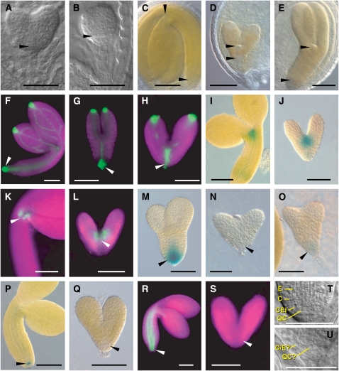
Figure 5.
Phenotype and Expression Patterns of Wild-Type and Homozygous rpn5a Embryos.
For each set of embryos, the wild type and mutant are shown at approximately the same developmental age. In all cases, the wild-type embryos are from heterozygous siliques of the same line as the corresponding mutant. In the fluorescent microscopy images, the tissue is highlighted by chlorophyll autofluorescence (false-colored magenta). In (A) and (B), arrowheads point to the abnormal shape of the cells in the position of the QC in rpn5a-1. In (C) to (U), arrowheads locate the RAM and SAM in the wild-type embryos or the equivalent position in the mutants. C, cortex; E, endodermis; QC?, cells in the QC position; C/E?, single cell file of unknown identity in the position of the cortex/endodermis. Bars = 50 μm.
(A) and (B) Young embryos of the wild type (A) and the rpn5a-4 mutant (B) at the heart stage.
(C) Mature embryos of the wild type and the rpn5a mutants.
(D) Example of the short embryo phenotype (rpn5a-2).
(E) Example of a long embryo phenotype (rpn5a-1).
(F) to (H) DR5rev:GFP expression in the wild type at the bent cotyledon stage (F) and rpn5a-4 mutant embryos with normal expression (G) and high expression that extended apically (H).
(I) and (J) STMpro:GUS expression in the wild type at the bent cotyledon stage (I) and rpn5a-4 (J).
(K) and (L) CUC1 (M0233 line) expression in the wild type at the bent cotyledon stage (K) and rpn5a-4 (L).
(M) to (O) PIN4pro:GUS expression in the wild type at the torpedo stage (M) and rpn5a-4 with no expression (N) or weak expression (O).
(P) and (Q) QC25pro:GUS expression in the wild type at the bent cotyledon stage (P) and rpn5a-4 (Q).
(R) and (S) SCRpro:GFP expression in the wild type at the bent cotyledon stage (R) and rpn5a-4 (S).
(T) and (U) Close-up of the root meristem in the wild type at the late heart stage (T) and rpn5a-3 (U) embryos.

