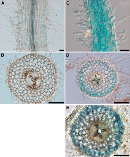
Figure 4.
Spatial Expression Pattern of the Srlk Promoter.
Histochemical localization of GUS activity in M. truncatula transgenic roots expressing the Srlk promoter:GUS fusion. Plants in (C) to (E) were treated for 6 h with salt (150 mM NaCl) before GUS staining. Bars = 250 μm in (A) and (C) and 500 μm in (B), (D), and (E).
(A) and (B) Whole-mount staining (A) of untreated roots allowed weak detection of blue staining in the epidermis that is barely detected in transverse sections (B).
(C) Whole-mount staining of NaCl-treated roots revealed strong staining of the epidermis and root hairs.
(D) and (E) Transverse sections of the transgenic roots after NaCl treatment showing strong blue staining, representative of GUS activity, in the root epidermis. In half of the cases, weak staining was observed in vascular tissues (E). GUS staining data are representative of at least 25 independent transgenic roots.

