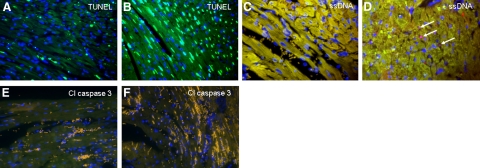
Figure 3.
Sections from a mouse heart undergoing 1 h of transient ligation and 24 h of reperfusion (A, C, E) and from a mouse heart subjected to 24 h of persistent ligation (B, D, F) exhibit positive TUNEL staining (A, B) (green) that is much in excess and is not congruent with positive ssDNA (C, D) (pink) or positive cleaved caspase 3 staining (E, F) (green, none seen). Arrows indicate rare cells positive for ssDNA. Nuclei were stained with DAPI (blue). Yellow color is attributable to autofluorescence of tissue; mustard color is attributable to red blood cells. All panels ×40.

