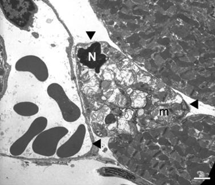
Figure 4.
Transmission electron micrograph from the peri-infarct zone in the heart from an animal subjected to 4 h of transient ligation and 24 h of reperfusion. Cardiomyocyte in the middle (arrowheads) displays morphology potentially consistent with apoptosis in this cell type, including a shrunken nucleus (N) with condensed chromatin and abnormal swollen mitochondria (m) with “wrinkled” bodies. Scale bar = 2 μm.

