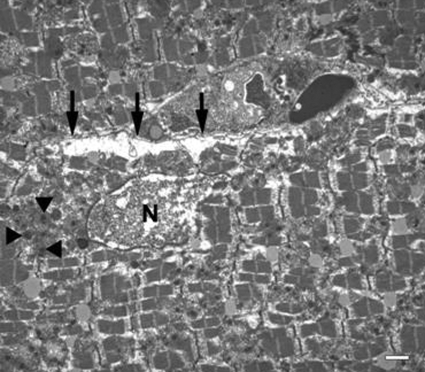
Figure 6.
Transmission electron micrograph from the infarct zone in the heart from an animal subjected to 4 h transient coronary ligation and 24 h of reperfusion. Note the classic morphological features of necrosis rather than apoptosis: disrupted nucleus (N) with loss of nuclear content, electron-dense floccular deposits in mitochondria (arrowheads), and ruptured plasma membrane (arrows). Scale bar = 1 μm.

