
Figure 3.
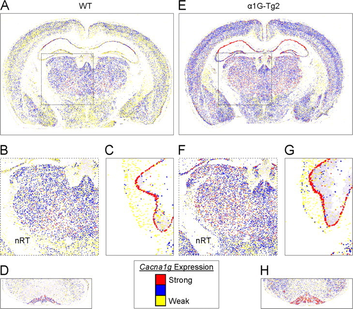
Accurate localization of α1G mRNA within the brain. A–H, Coronal brain sections from WT (A–D) and the higher-expressing α1G-Tg2 line (E–H) were hybridized with an α1G-specific digoxigenin-labeled riboprobe. Pseudocolored (“heat map”) images were generated by converting cellular Cacna1g mRNA expression intensity (see Materials and Methods and the in-figure legend), and they represent the midbrain (A, E) along with selected regions of interest, emphasizing the similar pattern but more intense hybridization signal indicated by color changes within the thalamus (B, F), Purkinje cell layer (C, G), and the cerebellar inferior olive (D, H). Note that expression of Cacna1g in the nRT is spared in the WT and transgenic mouse.