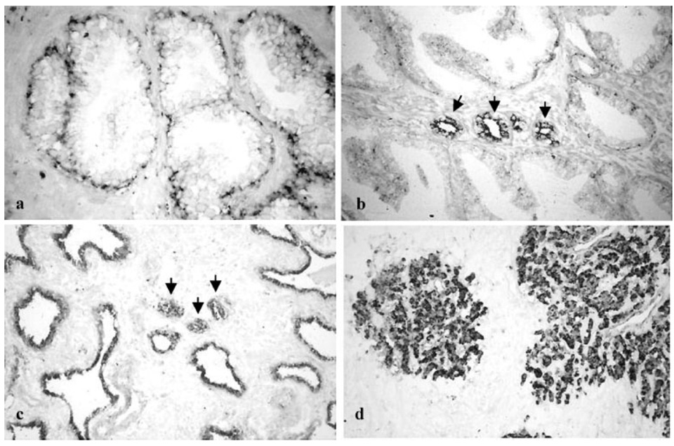
Fig. 2.
In situ localization of PCDH-PC m RNA in prostatic tissues. In situ hybridization technique was performed on formalin fixed paraffin-embedded tissue using digoxigenin-labeled PCDH-PC antisense probes, a: Tissues from normal prostate. Note the staining corresponding to PCDH-PC mRNA was mainly localized in the basal epithelial cells. Differentiated glandular cells were faint or negative-stained. Representative results of ISH performed on primary (untreated) cancers were presented in (b). Tumor cells (indicated by arrows) were strongly positive for PCDH-PC staining compared to adjacent normal epithelial cells. In tissues obtained from patients treated by hormonal therapy (c) and from hormone-refractory human CaPs (d), strong staining corresponding to PCDH-PC mRNA was localized in all tumor cells (indicated by arrows) and in normal (atrophic) epithelial cells. Magnification: a–d 200×.

