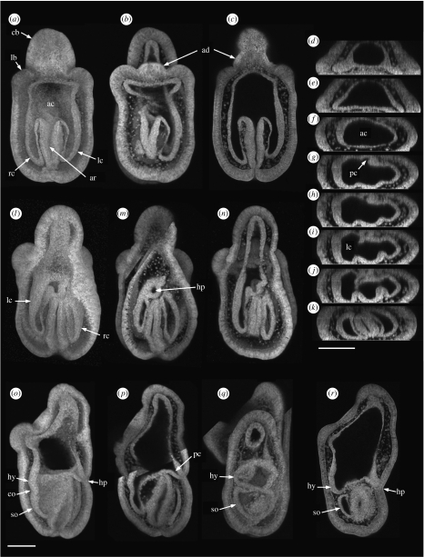
Figure 1.
Confocal images of the coelom in early brachiolaria larvae constructed from the Z stacks of hatched larvae (described in text). (a–c) Ventral views of one larva: (a) three-dimensional solid image, (b) three-dimensional transparent image, (c) single section from the Z stack. (d–k) Single transverse sections of the larva in (a–c) viewed from the larval posterior with dorsal uppermost. (l–n) Dorsal views of one larva: (l) three-dimensional solid image, (m) three-dimensional transparent image, (n) three-dimensional transparent image with more images cut from the front of the Z stack than in (m). (o,p) Lateral views of one larva, dorsal right: (o) three-dimensional solid image, (p) three-dimensional transparent image. (q,r) Lateral views of two larvae, dorsal right: (q) three-dimensional transparent image, (r) single section from the Z stack. (a–p) Four-days larvae; (q,r) 4.5-days larvae. Scale bars, 100 μm. ac, anterior coelom; ad, adhesive disc; ar, archenteron; cb, central brachium; co, constriction; hp, hydropore; hy, hydrocoele; lb, lateral brachium; lc, left lateral coelom; pc, pore canal; rc, right lateral coelom; so, somatocoele.

