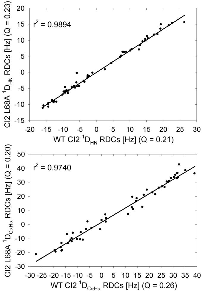
Figure 7.

A comparison of residual dipolar couplings (RDCs) for CI2 L68A versus WT CI2. Mutant N-H (top panel) and Cα-Hα (bottom panel) RDCs correlate extremely well with WT values, indicating that no significant structural rearrangements occur upon mutation. Q-factors for all variants were calculated using the WT CI2 structure. The correlations observed here are highly representative of the mutant/WT RDC comparisons for all mutants tested.
