Figure 2.
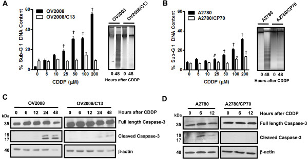
Sub-G1 DNA Content, DNA fragmentation, and cleavage of caspase-3 in OV2008, OV2008/C13, A2780 and A2780/CP70 cells treated with cisplatin. (A, left panel) OV2008 and OV2008/C13 cells were treated with or without the depicted concentrations of cisplatin for 1 h. Seventy-two hours later, floating and adherent cells were collected, fixed with 4% paraformaldehyde, stained with propidium iodide, and analyzed by microcytometric analysis using cell cycle software. (A, right panel) In a similar experiment, cells were exposed to a fixed 100 μM cisplatin concentration for 1 h and were harvested 48 h later; genomic DNA was isolated, separated by electrophoresis on a 2% agarose gel, impregnated with SYBR Gold nucleic acid stain, examined with an ultraviolet transilluminator, and photographed. (B) This graph depicts results from experiments similar to those shown in (A), but using A2780 and A2780/CP70 cells instead. *p < 0.05, #p < 0.01 and †p < 0.001 vs. control (0 μM cisplatin) for the respective cell line. (C and D) Cells were harvested at the indicated times after 1-h exposure to 100 μM cisplatin, whole cell proteins were isolated, equivalent amounts of proteins were electrophoresed, transferred to a PVDF membrane, and exposed to anti-caspase-3 or β-actin antibodies.

