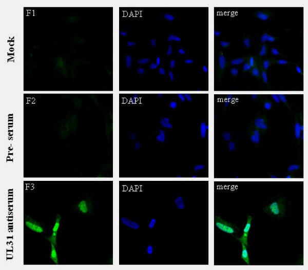
Figure 6.
Intracellular location of DEV UL31 protein analyzed by indirect immunofluorescence. Mock and DEV-infected cells were fixed with 4% formaldehyde at 36 h (p.i.) and processed for indirect immunofluorescence. Mock-infected cells with UL31 antiserum (F1). DEV-infected cells with preimmune sera (F2) or UL31 antiserum (F3). The cell nuclei were visualized by DAPI. (Images were acquired by using 40× objective)

