Fig. 4.
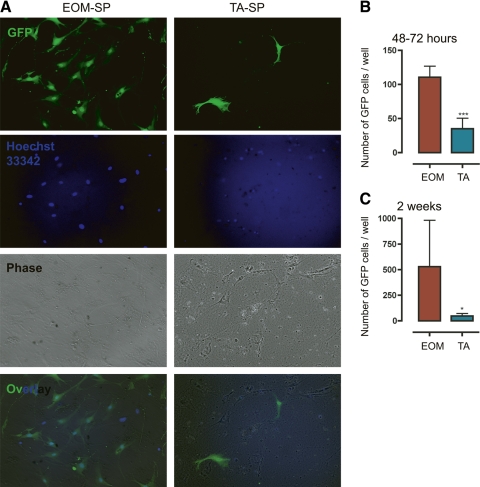
EOM-SP cells proliferate better in vitro than TA-SP cells. A: representative culture of EOM and TA green fluorescent protein-positive (GFP+) SP cells after 2 wk of culture over a myoblast feeding layer. Equal numbers of GFP+ SP cells were plated on a mdx-myoblast feeding layer. After 18 days, cells were counted. Left: EOM-SP. Right: TA-SP. From top to bottom: GFP, Hoechst, phase, and overlaid (Ov) images. B: a higher number of EOM-SP cells than TA-SP cells was present in culture 48–72 h after sorting. Cells were counted with a fluorescence microscope. Means ± SD are shown; n = 2. ***P < 0.0001. C: differences in the number of EOM and TA GFP+ cells in cultures increased over time. After 13–18 days, cells were counted with a fluorescence microscope. Means ± SD are shown; n = 3. *P < 0.05.

