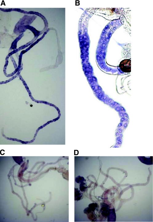
Fig. 4.
In situ hybridizations for CG2196 in Malpighian tubules of adult Oregon R flies. Staining with anti-sense probe. A and B: clear staining appears in the ureter and in the principal cells of the main and the lower segment of both the posterior and anterior Malpighian tubules. In most cases, the staining in the main segment of the tubule was stronger compared with that of the lower segment. The initial segment was not stained in both pairs of tubules. C and D: sense controls. In all photographs, the diameter of the tubule can be taken as 35 μm.

