
Figure 4.
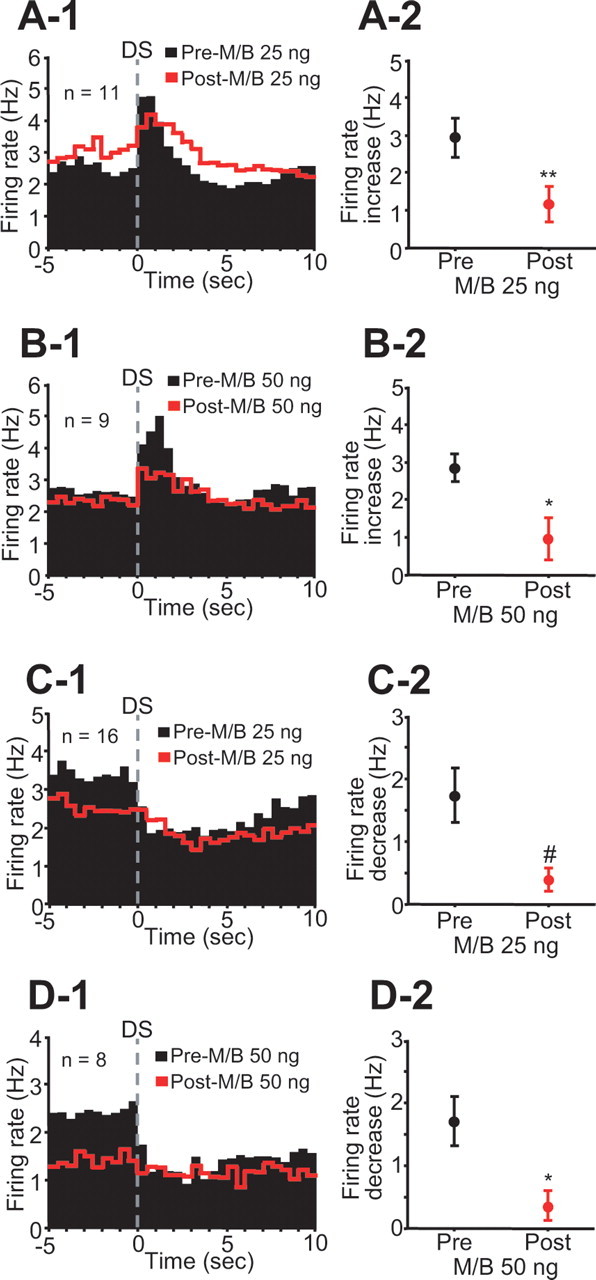
Inactivation of the dmPFC reduces incentive cue excitation and inhibition on trials when the animal makes a behavioral response to the DS. A-1, B-1, Averaged PETHs (0.5 s bin width) of DS-excited neurons before (black) and after (red) 25 and 50 ng M/B injections. A-2, B-2, Comparisons of DS excitation between preinjection and postinjection of each drug. C-1, D-1, Averaged PETHs (0.5 s bin width) of DS-inhibited neurons before and after each drug injection. C-2, D-2, Comparisons of DS inhibition between preinjection and postinjection. #p < 0.05, *p < 0.01, **p < 0.001 compared with the preinjection epoch.