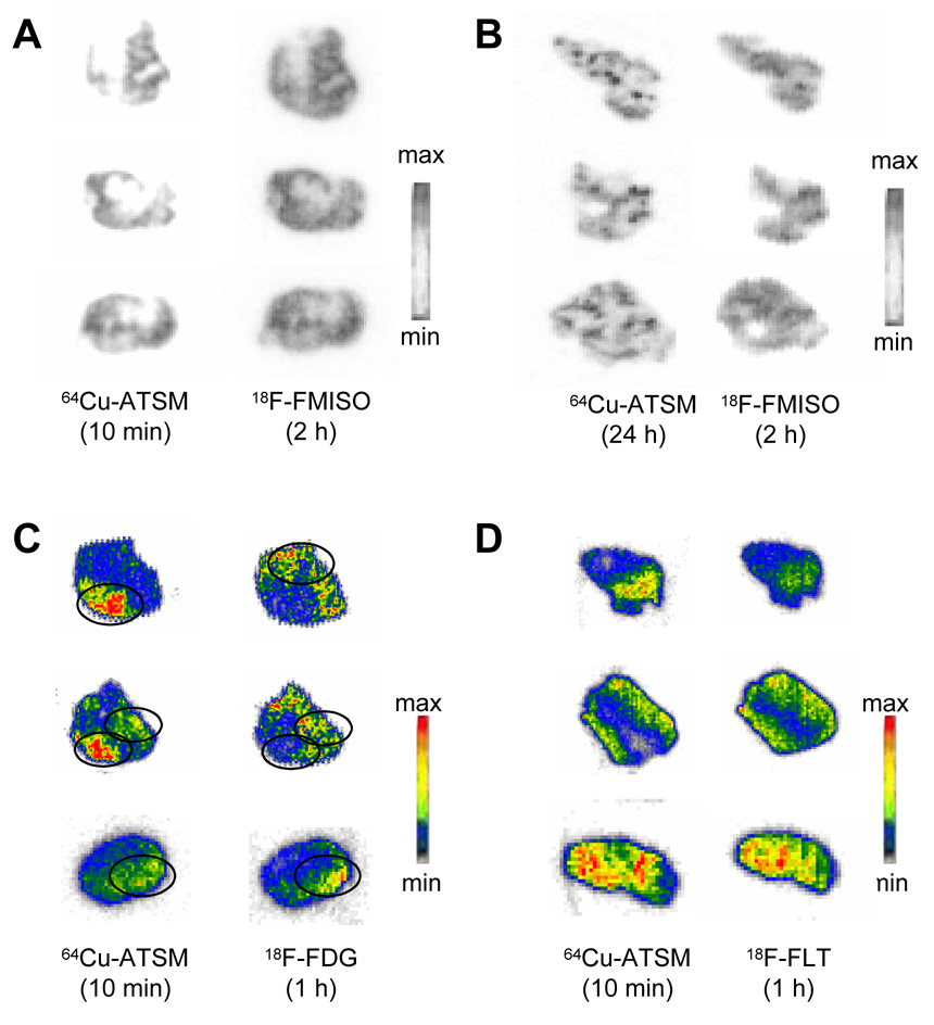
Fig. 1.
Ex vivo electronic autoradiographs of the regional in vivo uptake of (A, B) 18F-FMISO, (C) 18F-FDG, (D) 18F-FLT with 64Cu(ATSM) in 9L gliosarcomas. The images shown are representative of the typical dual-tracer autoradiographs obtained from all of the slices of the 9L tumors from rats. Shown in each panel are three representative slices chosen at random from a total of 120 slices from 5 tumors. Shown are slices with following 18F tracer collection (right) and the 64Cu-ATSM distribution (left) in the same tumor slices. At the time of imaging, the 18F images are generated 99% by 18F. Following 24 h decay the images are generated from 95+% 64Cu.

