Fig. 1.

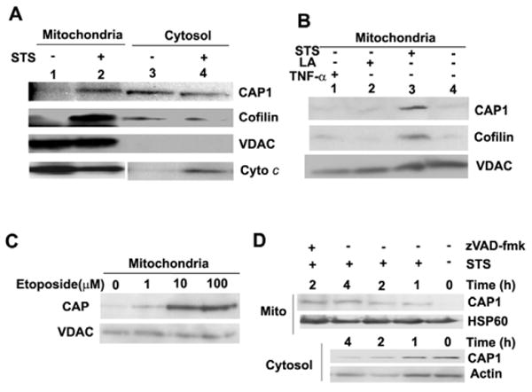
CAP1 translocates to mitochondria upon treatment to induce apoptosis. (A) STS stimulates CAP1 translocation to mitochondria. HeLa cells were treated with 1 μM STS for 4 hours, mitochondria were isolated by sucrose gradient centrifugation and mitochondria or cytosol fractions were subjected to western blot. Cytochrome c (Cyto c) images were from the same gel, although mitochondrial and cytosol results were scanned separately because a longer exposure was required to detect signals in the cytosol. (B) STS but not LA stimulated translocation of CAP1 to mitochondria. HeLa cells were treated with STS, LA and TNFα, mitochondria were isolated as above and CAP1 and cofilin were detected. (C) Treatment with etoposide also induces CAP1 translocation to mitochondria. HeLa cells were treated for 24 hours with etoposide concentrations ranging from 1 μM to 100 μM, mitochondria were purified and subjected to western blots with CAP1. (D) Time course of CAP1 translocation to the mitochondria. HeLa cells were treated with STS (1 μM) in the presence of 50 μM zVAD-fmk, where indicated. Gradient-purified mitochondrial (top) and cytosolic (bottom) samples were subjected to western blots to detect CAP1. HSP60 and actin were used as loading standards.
