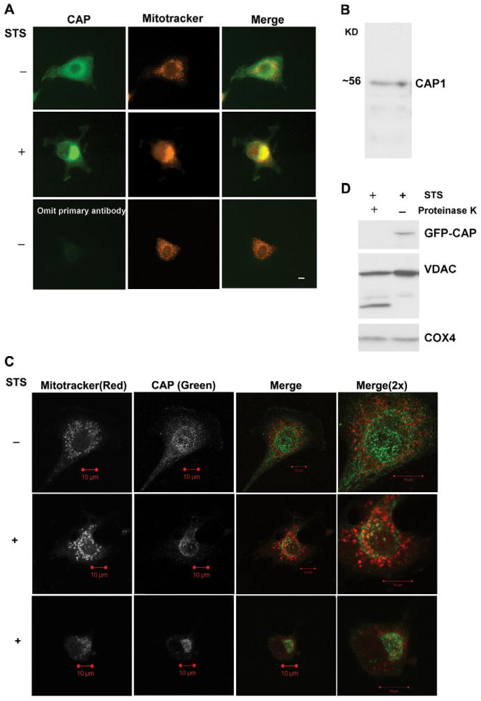
Fig. 2.

CAP1 translocates to the outer mitochondrial membrane. (A) CAP1 translocates to mitochondria upon apoptosis induction with STS in NIH3T3 cells. Cells were plated on coverslips overnight and stained with 50 nM MitoTracker Red CMXROS for 1 hour followed by treatment for 1 hour with (+) or without (−) 1 μM STS. Fixed and permeabilized cells were incubated with mouse anti-CAP1 antibody for 2 hours and then incubated for 1 hour with anti-mouse FITC conjugate. The slides were analyzed using a 40× objective lens by fluorescence microscopy. Scale bar: 10 μm. (B) Western blots of NIH 3T3 cell lysate with anti-CAP1 antibody demonstrate specificity for the antibody. (C) Confocal imaging of CAP1 reveals punctate staining at the mitochondria in treated cells. NIH3T3 cells were treated and processed as described above. Fluorescence images were collected at similar focal planes by confocal microscopy with a 63× objective lens; two independent STS-treated cells are shown. Scale bars: 10 μm. (D) Mitochondrial CAP1 is proteinase sensitive. HEK293T cells were transfected with GFP-CAP1 plasmid and treated with STS for 2 hours. Gradient-purified mitochondria were digested with proteinase K as indicated and then subjected to western blot analysis.
