
Fig. 4.
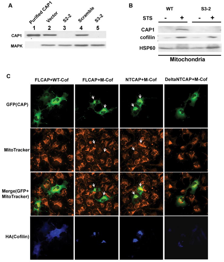
Knockdown of CAP1 and its interaction with cofilin. (A) CAP1 knockdown in HeLa cells. Stable cell lines S2-2 and S3-2 are from two independent shRNA constructs, S2 and S3, which target different sequences within CAP1 (see Materials and Methods). Control cell lines include HeLa cells harboring empty vector or a scrambled shRNA S2. Purified CAP1 from pig platelet was used as a positive control and MAPK was blotted to normalize total protein. CAP1 levels in the cell lysates were detected by western blotting. (B) CAP1 knockdown does not affect cofilin translocation to mitochondria during apoptosis induction. HeLa wild-type or CAP1-knockdown cells (S3-2) were treated with 1 μM STS for 2 hours. Sucrose-gradient-purified mitochondria were isolated and analyzed by western blot. HSP60 was monitored to normalize total mitochondrial protein. (C) Mitochondrial cofilin stimulates CAP1 translocation to mitochondria. GFP-tagged CAP1 constructs, FL-CAP, NT-CAP and ΔNTCAP1 were cotransfected with mitochondria-targeted cofilin (M-cof) into COS-7 cells and stained with MitoTracker. Cofilin was stained with anti-HA monoclonal antibody 12CA5. Images were viewed on a fluorescence microscope with a 40× objective lens. Arrows indicate cells with localization of GFP fusion CAP1 (domains) to mitochondria.