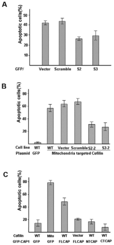
Fig. 5.

CAP1 interacts with cofilin to promote apoptosis. (A) Transient expression of CAP1 shRNA in HeLa cells reduces STS-induced apoptosis. HeLa control and CAP1-knockdown cell lines were cotransfected with GFP and ShRNA constructs (1:1). After 24 hours, cells were treated with 1 μM STS for 2 hours and labeled with Hoechst 33342. GFP-positive cells were counted. Cells with DNA fragmentation and nuclear collapse were scored as apoptotic cells. ∼200 cells were counted for each treatment and the experiment was done three times with similar results. Percentage of apoptotic cells are shown as mean ± s.e.m. (B) CAP1 knockdown reduces cofilin-induced apoptosis. CAP1-knockdown HeLa cells were transfected with mitochondrial cofilin along with GFP. 24 hours after transfection, cells were stained with Hoechst 33342 and scored for chromosome condensation. Approximately 100 transfected cells were scored for each of three fields and the experiment was repeated three times with similar results. Data are presented as mean ± s.e.m. (C) Overexpression of CAP1 stimulates cofilin-induced apoptosis. HEK293T cells were cotransfected with GFP-fused FL-CAP1, NT-CAP1 or CT-CAP1 along with either M-cofilin or wild-type cofilin (0.4 μg and 0.8 μg, respectively, per well). Cells were allowed to express proteins for 18 hours and then fixed cells were stained with 1 μg/ml Hoechst 33342 for 10 minutes. Apoptosis was identified by nuclear condensation and DNA fragmentation under fluorescence microscopy in GFP-positive cells. At least 300 GFP-positive cells were scored for each of three fields and experiment was repeated three times with similar results. Data are presented as mean ± s.e.m.
