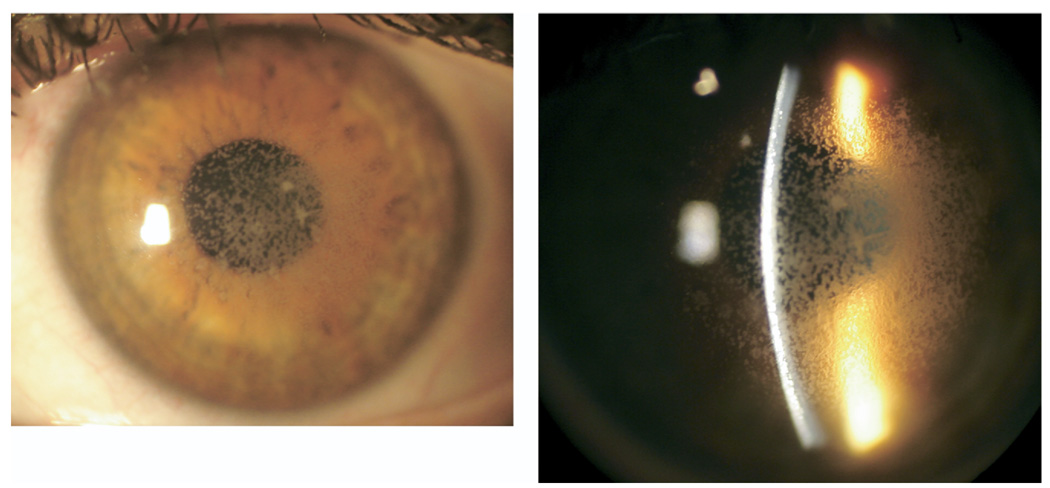
FIGURE 1.
Avellino corneal dystrophy worsening after laser in situ keratomileusis (LASIK). (Left) Slit-lamp photography of the cornea of the left eye showing extensive granular deposits. (Right) The deposits are scattered in the anterior stroma, but are mainly concentrated in the LASIK flap interface.

