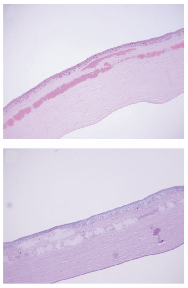
FIGURE 2.
Avellino corneal dystrophy worsening after LASIK: histopathologic findings. (Top) Histologic section of the corneal recipient tissue (stain, hematoxylin and eosin; original magnification ×200). The deposits are seen in the sub-Bowman layer, anterior stroma, and the LASIK interface. The size of the deposits tends to increase as they approach the interface. (Bottom) Periodic Acid-Schiff section, with the deposits failing to stain (original magnification ×200).

