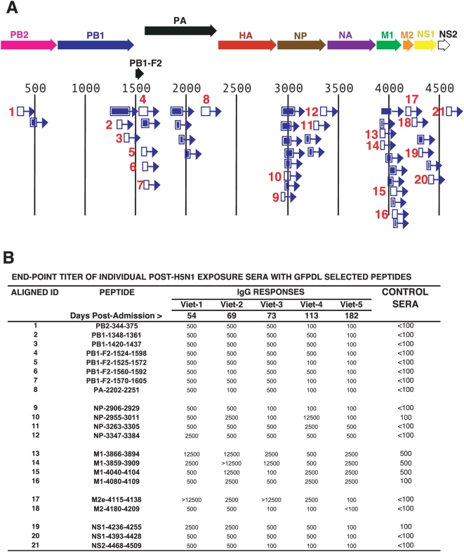
Figure 5. Antibody epitopes in H5N1 internal proteins (FLU-6) recognized by pooled sera from H5N1 infected individuals.
(A) Schematic alignment of the peptides identified using GFPDL (H5N1-FLU-6) expressing all internal proteins of influenza A/Vietnam/1203/2004. The predicted influenza encoded proteins are numbered according to the complete proteome (Figure S1). Bars with arrows indicate identified inserts in the 5′–3′ orientation. Numbered segments represent high frequency clones (≥5; Table S1). These peptides were expressed and purified from E. coli or were chemically synthesized and the numbers correspond to the peptide identifiers in the ELISA assay in (B). (B) Reactivities of sera from individual H5N1-infected patients (Viet 1–5) or sera from healthy Vietnamese adults against peptides derived from: PB2 (1); PB1 (2–3); PB1-F2 (4–7); PA (8); NP (9–12); M1 (13–16); M2e (17); M2 (18); NS1 (19–20); and NS2 (21).

