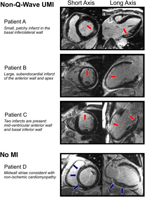
Figure 1. Typical DE-CMR images.
Short and long axis views of DE-CMR images from four patients are shown. Patients A–C demonstrate hyperenhancement (red arrows) consistent with prior myocardial infarction. None had Q-waves on electrocardiography, and all three were classified as having non-Q-wave UMI. Of note, Patient C has evidence of two distinct infarcts. Patient D has hyperenhancement (blue arrows) involving the midwall of the interventricular septum, sparing the subendocardium. This pattern is not consistent with prior myocardial infarction, and this patient was categorized in the “no MI” group. See text for further details.

