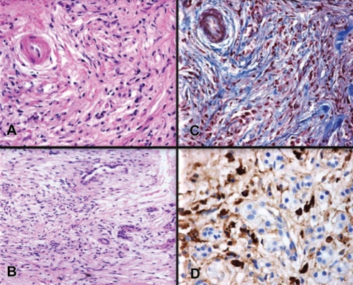
Figure 1).
Pancreatic histology of a 52-year-old woman with autoimmune pancreatitis. A Duct showing typical periductal lymphoplasmacytic inflammation and narrowing of the lumen. B Lymphoplasmacytic infiltration. C Periductal and interlobular fibrosis. D Immunohistochemical staining for immunoglobulin G4 showing marked immunoglobulin G4-positive plasma cell infiltrates. Courtesy of Dr Luis Uscanga, Teaching Department, INCMNSZ, Mexico City, Mexico

