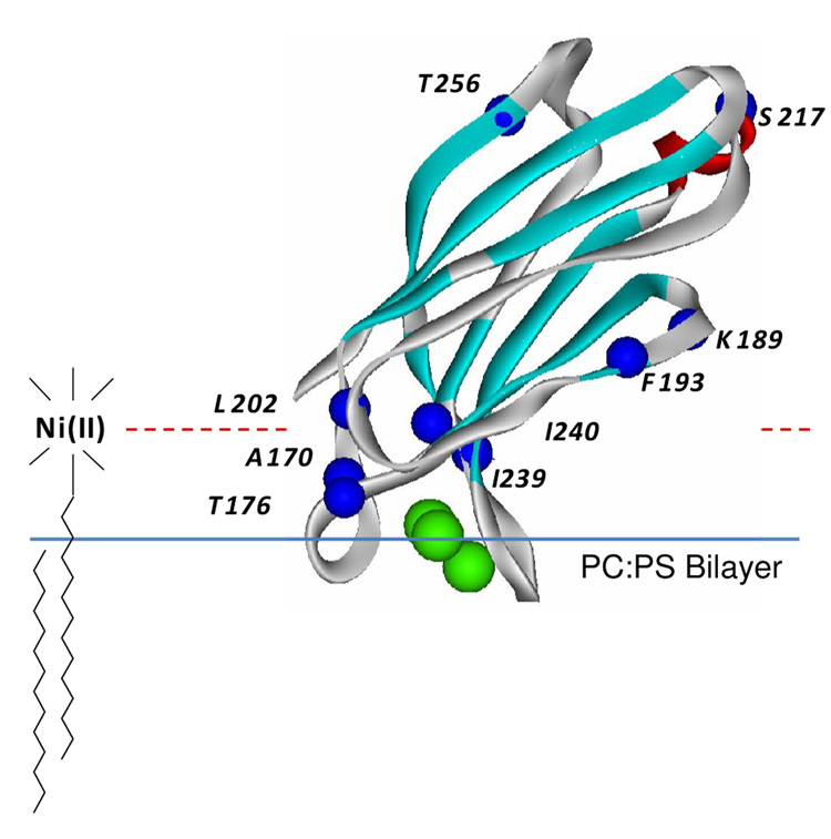
Figure 5.
High resolution model for syt1C2A (PDB 1BYN) (15). The domain is shown bound to bilayers composed of POPC:POPS with a depth of penetration and orientation that were previously determined from SDSL (28). This structure was used to calibrate the position parameter Φ2 as described in Methods. This parameter is expected to be sensitive to the position of the label on the aqueous side of the bilayer interface and utilizes the lipid bound nickel chelate, DOGS-NTA-Ni(II). Single cysteine mutants were generated at the indicated residues (Cα carbons highlighted in blue) to incorporate a series of R1 labels (see Fig. 1A) at varied positions off the bilayer interface.

