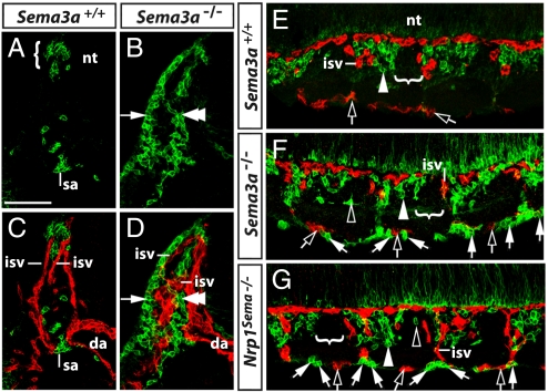
Fig. 4.
Relationship of ectopic NCCs to blood vessels. Transverse (A–D) and longitudinal sections (E–G) stained for p75 (green) and endomucin (red) illustrate the relationship of NCCs to blood vessels at 9.5 dpc. (A and B) At the level of the intersomitic furrow, NCCs in wild-types accumulated in the MSA (bracket in A), whereas NCCs in Sema3a-null mutants formed a ventromedial (double arrowhead) and dorsolateral (arrow) stream that followed the course of intersomitic blood vessels (C and D). (E) Longitudinal sections demonstrated that most NCCs normally migrated into the anterior sclerotome (white arrowhead), but not into the posterior sclerotome (indicated with a bracket). (F and G) In Sema3a- and Nrp1Sema-null mutants, only few NCCs entered the anterior sclerotome (white arrowhead), while most NCCs (white arrows) migrated alongside intersomitic and perisomitic vessels (clear arrows). In addition, ectopic NCCs were occasionally seen in the posterior sclerotome of mutants (clear arrowheads). Abbreviations: da, dorsal aorta; isv, intersomitic vessel; nt, neural tube; sa, sympathetic anlage. (Scale bar, 100 μm.)

