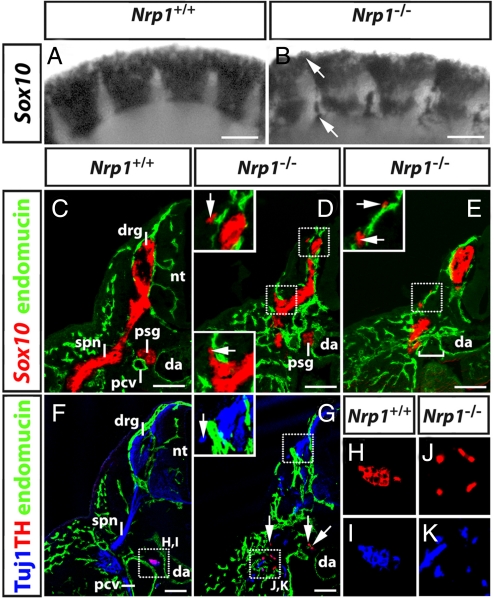
Fig. 5.
Ectopic NCC migration impacts on PNS organization. (A and B) Wholemount in situ hybridization of 9.75 dpc wild-type and Nrp1-null embryos for Sox10 revealed ectopic NCC near the dorsal neural tube and in the intersomitic furrows (arrows). (C–E) Transverse sections of 10.5 dpc wild-type and Nrp1-null embryos labeled for Sox10 (red) and endomucin (green); half of each section is shown. (C) In wild-types, primary sympathetic ganglia (psg) formed between the dorsal aorta (da) and the posterior cardinal veins (pcv), while dorsal root ganglia (drg) form adjacent to the neural tube (nt) and extended 1 spinal nerve each (spn). (D and E) In mutants, dorsal root ganglia and primary sympathetic ganglia formed, but they were smaller than normal; at some axial levels, the sympathetic ganglia were missing (square bracket in E). The insets in D and E are higher magnifications of the squared areas and illustrate the proximity of ectopic cell clusters to superficial blood vessels. (F and G) Transverse sections of 10.5 dpc wild-type and Nrp1-null embryos labeled for neuronal microtubules (Tuj1; blue), endomucin (green), and the sympathetic marker tyrosine hydroxylase (TH; red). The inset in G is a higher magnification of the squared area and illustrates the presence of ectopic neuronal microtubule-positive, TH-negative neurons adjacent to the dorsal root ganglia. In mutants, TH-positive neuronal progenitors were scattered distally to the dorsal aorta. Ventral regions marked with squares in F and G are shown as separate color channels at higher magnification in H and I and J and K, respectively. (Scale bars, 100 μm.)

