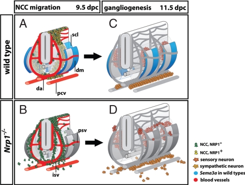
Fig. 7.
Working model: SEMA3A and NRP1 control trunk NCC migration to organize PNS neurons. (A and B) NCC migration pathways at 9.5 dpc. (A) In wild-types, only a few intermediate wave NCCs are NRP1-negative (green) and travel alongside intersomitic and perisomitic vessels (red). Rather, most NCCs are NRP1-positive (yellow) and are channeled into the anterior sclerotome by repulsive SEMA3A signals. Accordingly, Sema3A (blue) is expressed in 2 domains, a narrow stripe in the dermomyotome adjacent to the preceding intersomitic furrow, and a broader domain in the posterior dermomyotome that extends into the posterior sclerotome. (B) In the absence of NRP1 signaling, intermediate wave NCCs are blind to SEMA3A (now shown in gray) and preferentially migrate alongside intersomitic blood vessels (red), similar to early wave NCCs. (C and D) Peripheral neuron position in wild-types and Nrp1-null mutants at 11.5 dpc reflects the migratory patterns of their NCC precursors. (C) In wild-types, sensory neurons condense into segmentally arranged dorsal root ganglia in the anterior sclerotome of the somites, while sympathetic neurons form paired, but nonsegmented primary sympathetic cords next to the dorsal aorta. (D) In the absence of SEMA3A/NRP1 signaling, sensory and sympathetic neurons differentiate in ectopic positions corresponding to the earlier position of their NCC precursors. Consequently, both the segmentation of the sensory system and the assembly of the sympathetic cords are disrupted. Abbreviations: da, dorsal aorta; dm, dermomyotome; isv, intersomitic vessel; psv, perisomitic vessel; pcv, posterior cardinal vein; scl, sclerotome.

