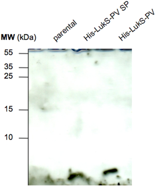
Figure 9. The signal peptide is present in cell wall associated material.
The 2% SDS extract of various strains were run on 17% SDS PAGE, transferred to nylon membrane and probed with mouse monoclonal anti-poly His antibody (1∶500) (Sigma) followed by goat anti mouse HRP conjugated antibody (1∶2000) (Sigma). Parental (LUG960) indicates the genetic background in which the genetic modifications were made; His-LukS-PV SP, parental carrying a plasmid encoding the His-tagged LukS-PV signal peptide alone; His-LukS-PV, parental carrying a plasmid encoding the His-tagged full length LukS-PV.

