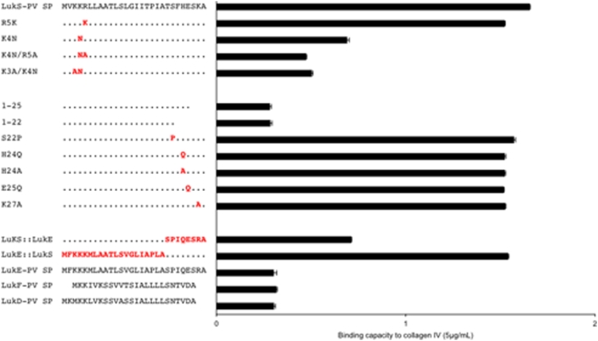
Figure 12. Identification of LukS-PV signal peptide residues involved in the adhesion phenotype using mutagenesis.
Sequences of wild type LukS-PV signal peptide (top) and mutants are shown on the left side, together with the alignment of sequences of LukE, LukF-PV and LukD signal peptides (bottom). The binding capacity of the corresponding sequences to type IV collagen (5 µg/mL, Sigma) is shown on the right side. Mutated amino acids are colored red while other LukS-PV SP amino acids are represented by dots. Deletion mutants of LukS-PV SP are denoted by their sequence length (1–25 and 1–22).

