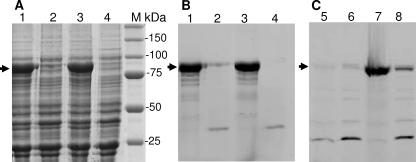
FIGURE 4.
Expression and fluoresceyl-ampicillin labeling of some PBP1b mutants. A and B, Coomassie Blue staining (A) and fluoresceyl-ampicillin (50 μm) labeling (B) of PBPs after SDS-PAGE loaded with equivalent amount of membrane extract from cells expressing PBP1b mutants at 37 °C. Lane 1, F237A; lane 2, T267A; lane 3, Q271A; lane 4, R286A; M = molecular mass standard. C, fluorescence detection of fluoresceyl-ampicillin labeled PBPs at 30 °C. In lanes 5 and 6, the expression of the PBP1b R286A was realized at 18 and 37 °C, respectively. In lanes 7 and 8, the expression of PBP1b T267A was realized at 18 and 37 °C, respectively (no overexpression of R286A was visible after Coomassie Blue staining, not shown).

