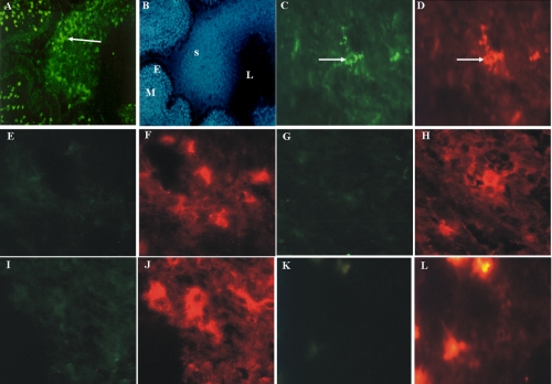
FIGURE 1.
Human tissue eosinophils from infected CF patients contain exclusively 3NT-positive residues. A and B, fluorescence micrograph of 3NT-positive cells in the lung mucosa (panel B, M) and in the sputum (panel B, S) filled bronchial lumen (panel B, L) of a patient with CF stained with a rabbit NT antibody (A) and DAPI (B)(panel B, E (epithelium)), original magnification: A and B, ×100. Double fluorescence micrographs of CF tissue sections stained with a rabbit antibody against 3NT (panels C, E, G, I, and K) and markers specific for eosinophils (D), T-lymphocytes (F), mast cells (H), monocytes (J), and neutrophils (L). Only eosinophils were 3NT-positive (panels C and D, arrows). Original magnification: panels C-L, ×400.

