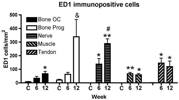
Figure 3.
Mean (+ SEM) ED1-positive cells (a marker of macrophages, osteoclasts (OC) or their progenitors (Prog)) was examined in distal radius and ulna bones, median nerves, forelimb flexor muscles or forelimb flexor tendons. Normal controls (C, n=4) and rats that had performed the MRHF task for 6 weeks (n=4) or 12 weeks (n=4) were evaluated. Significant increases from control levels are denoted by symbols (*p<0.01 and **p<0.001 compared to controls; # p<0.001 compared to bone osteoclasts, muscle or tendon).

