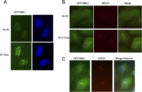
Figure 2.
NBA1 accumulates to DNA damage sites in response to IR. (A) NBA1 localizes to IRIF. U2OS cells that stably express a GFP-NBA1 fusion were treated with 10 Gy IR followed by a 4-h incubation at 37°C. Cells were then extracted with 0.5% Triton X-100, fixed with 3.7% formaldehyde, and stained with an anti-GFP antibody and Hoechst dye. (B) GFP-NBA1 forms IRIF that colocalize with BRCA1 foci. U2OS cells stably expressing GFP-NBA1 protein were treated with 10 Gy IR. After 2h incubation of cells post-IR, cells were fixed and stained with antibodies against GFP and BRCA1. (C) GFP-NBA1 accumulates to laser-induced DNA damage regions that colocalize with γ-H2AX. U2OS cells stably expressing GFP-NBA1 were treated with a laser for microirradiation.

