Figure 4.
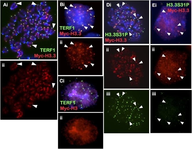
Cellular distribution of MYC-H3.3 and MYC-H3 in mouse ES cells. (A–C) MYC-H3.3 and MYC-H3 constructs were transfected into ES129.1 cells and examined for chromosomal localization 24 h post-induction with 1 μM doxycycline. Both N-terminal and C-terminal (data not shown) MYC-H3.3 was enriched along the chromosome arms and at the telomeres in metaphase (A, ii, red) and interphase (B, ii, red) (arrowheads show some examples) ES129.1 cells, as indicated by costaining with anti-TERF1 antibody (A,B, i, green). N-terminal (C, ii, red) and C-terminal (data not shown) MYC-H3 localized to all regions of the chromosomes. (D,E) MYC-H3.3 (ii, red) also showed colocalization with H3.3S31P in metaphase (D, iii, green) but not interphase (E, iii, green) ES129.1 cells.

