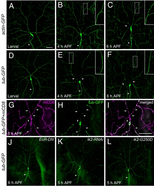
Fig. 3.
The cytoskeletons of ddaC neurons in wild type and mutants during dendrite pruning. (A–C) The actin cytoskeletons were visualized by anti-GFP antibody staining in wild-type animals with UAS-actin-GFP driven by ppk-GAL4. (A) The actin-GFP proteins of ddaC cells in larva. (B) At 4 h APF, the actin cytoskeletons in the proximal dendrites of ddaC showed signs of disruption, becoming thinner and blebbing. (C) Breakage of actin cytoskeletons (arrow) was evident at 6 h APF. (D–F) The microtubules in wild-type ddaC neurons, which expressed UAS-tub-GFP (tubulin-GFP) driven by ppk-GAL4 and labeled by anti-GFP antibodies, were intact in larva (D), and showed breakage in proximal dendrites (arrows) at 4 h APF (E) and 6 h APF (F). Magnified views (Insets) of the more distal dendrites (dashed boxes, ≥120 μm from the soma) showed that the actin cytoskeletons (B and C) and the microtubules (E and F) were fairly intact. (G–I) Double staining was carried out to visualize the membrane by mCD8 signals (G) and the microtubules by GFP signals (H) in wild-type ddaC neurons at 5 h APF. The proximal regions of dendrites showed the gaps of microtubules (arrows in H) and the presence of continuous membrane (arrows in G). The merged image was shown in I. (J–L) The microtubules revealed by the expression of ppk-GAL4, UAS-tub-GFP, and anti-GFP antibody staining were intact in the pupae with the expression of dominant negative ecdysone receptor (EcRDN-W650A) (J), of ik2-RNAi (K), and of dominant negative ik2 (ik2-G250D) (L). Arrowheads indicate the axons. (Scale bars, 50 μm.)

