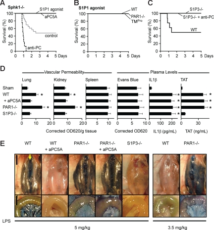
Figure 4.
Roles of S1P1 and S1P3 signaling in protection from LD50 LPS-induced lethality. (A) Sphk1−/− mice exhibit mortality reduction by aPC5A or S1P1 agonist AUY954 treatments and mortality enhancement following anti-PC TVM1 antibody treatment (n = 8–22 mice/group; P < .001, aPC5A, S1P1 agonist, and TVM1 treatment vs control). (B) Intervention at 10 hours with S1P1 agonist AUY954 rescued wild-type, PAR1−/−, and TMPro mice from death after LD50 challenge (n = 8/group; P < .02 vs respective control). (C) Protection of LD50-challenged S1P3−/− mice from anti-PC TVM1 antibody treatment (n = 8/group; P < .01 vs wild-type control). (D) Vascular leakage in lungs, kidney, and spleens, and plasma levels of Evans blue, IL1β, and TAT in LD50-challenged wild-type (WT), PAR1−/−, and S1P3−/− mice treated with aPC5A (mean ± SD; n = 3-7 mice/group; * indicates P < .01 different from sham receiving no LPS, ANOVA). (E) Macroscopic views at 18 hours after LPS of the anterior abdominal wall 45 minutes after Evans blue injection were prepared by blunt dissection of the superficial fascia. For orientation, the arrow points to the sternal xiphoid where the linea alba (running top to bottom) originates and the skin flap is turned to the left. The close-up views of the mesentery and small intestine (bottom panels) were taken after whole-body perfusion. Scale bars equal 1 cm for overviews and 5 mm for close-ups.

