Figure 3.
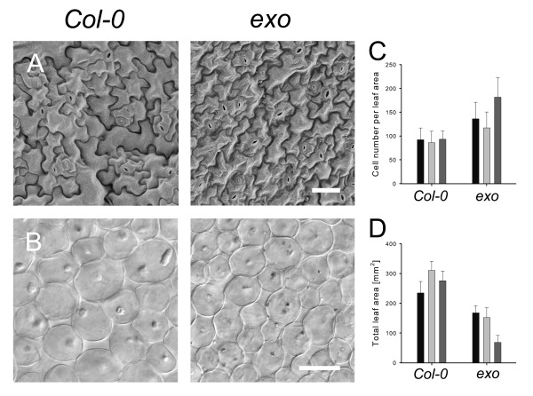
Cell size and leaf area of exo rosette leaves. Plants grown for 35 days in a greenhouse were subjected to microscopic analyses. A. Scanning electron micrograph (SEM) of abaxial epidermal cells of fully expanded 5th or 6th rosette leaf of wild-type and exo. SEM images have the same magnification, the bar represents 40 μm. B. Palisade cells in subepidermal layer of wild-type and exo. Bar represents 40 μm. C. Number of palisade cells in subepidermal layer per leaf area (144.000 μm2). Data of three independent experiments are shown (mean ± SD). 20 leaves per experiment were analyzed. D. Area of leaf blades. Data of three independent experiments are shown (mean ± SD).

