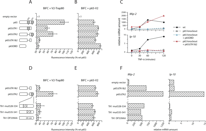
Figure 5. Mutations of p65 That Prevent Interaction with Trap-80.
(A) Deletion of TA1 and TA2 prevents the interaction of p65 with Trap-80.
(B) All p65 deletion mutants tested can still homodimerize.
(A, B, D, and E) depict fluorescence intensities of HEK-293 cells after co-transfection with vectors expressing the indicated mutants of p65 fused to Venus fragment 1 (V1), and either V2-Trap-80 (A and D) or p65-V2 (B and E). Intensities are expressed as a percentage of the level in cells co-expressing p65-V1 and V2-Trap-80 (shown in [A]). The residual ∼30% fluorescence seen with the p65 DBD in (A) is similar to the level seen in cells co-expressing V1 and V2 fused to control, noninteracting proteins. Error bars indicate standard errors of independent transfections. The results presented here are representative of two to four experiments.
(C) Deletion of TA1 and TA2 prevents expression of Ip-10 but does not affect expression of Mip-2. p65-knockout fibroblasts were transduced with retroviruses driving expression of the p65 DBD (blue triangles) or of p65ΔTA1&2 (red triangles), and expression of Ip-10 and Mip-2 was compared to wild-type and untransfected p65-knockout cells.
(D) Mutations in TA1 that prevent the interaction of p65ΔTA2 with Trap-80.
(E) All p65 mutants tested can still homodimerize.
(F) Mutants that do not interact with Trap-80 cannot drive expression of Ip-10, but expression of Mip-2 is unaffected. Expression of Mip-2 (left) and Ip-10 (right) mRNA in p65-knockout fibroblasts transduced with retroviruses expressing the indicated p65 mutants, after stimulation for 1 h with TNF-α.

