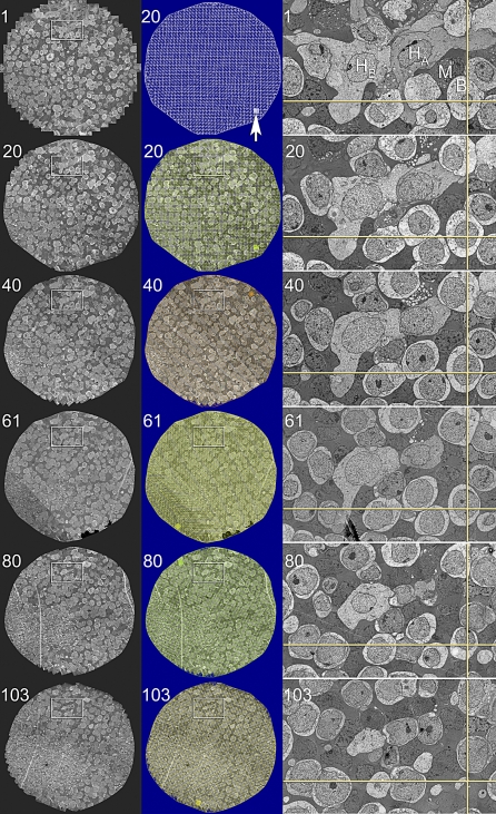
Figure 11. Automatic Registration of Canonical Scale Mosaics.
The left two columns are six 1,000+ tile mosaics from a series of over 120 horizontal plane 70-nm sections of the rabbit inner nuclear layer (sections 1, 20, 40, 61, 80, 103) spanning over 9 μm. Each mosaics is 250 μm wide. The middle column shows mosaics 20, 40, 61, 80, 103 with a colored overlay of the tile adjustment mesh (the true subtile mesh is much finer). The high contrast version of the mosaic 20 mesh shows that the bounding and bisecting lines only slight deviations from linearity due to slice-to-slice distortions. However, these do not accumulate. The arrow indicates a patch of the true mesh density. The right column (53 μm wide) is a magnified region of each slice showing the excellent cell-to-cell and subcellular alignment achieved by purely automatic image registration with ir-tools.

